


1. Introduction
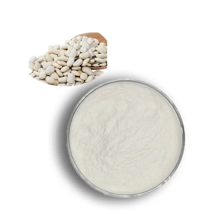
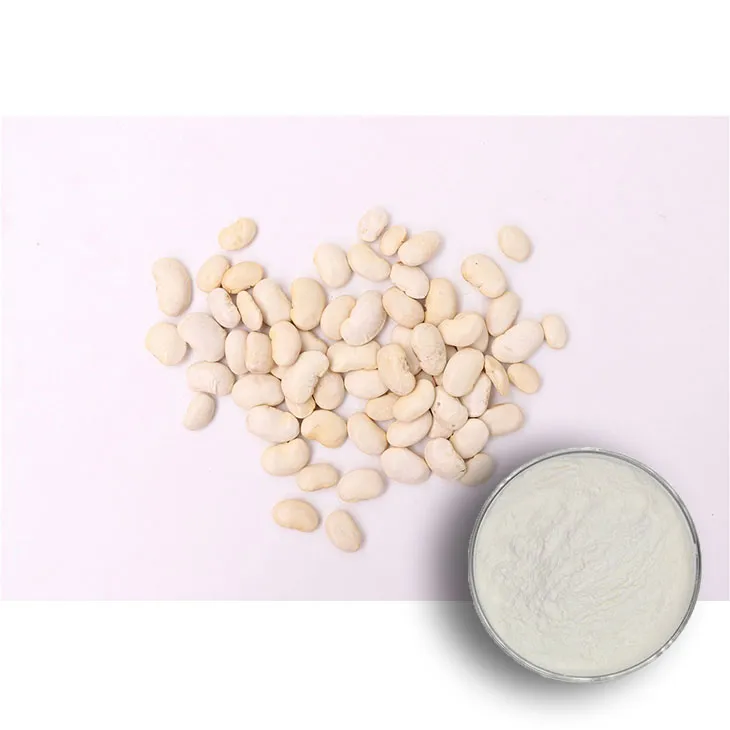
Kidney beans are a common and important source of plant proteins. Understanding how to effectively extract, separate, and identify proteins from Kidney Bean Extracts is crucial for various applications, including food science, nutrition research, and biotechnology. This article aims to provide a comprehensive overview of the processes involved in obtaining high - quality proteins from Kidney Bean Extracts.
2. Extraction of Proteins from Kidney Bean Extract
2.1. Sample Preparation
The first step in protein extraction is proper sample preparation. Kidney beans should be carefully selected, ensuring they are free from contaminants and damage. They are then washed thoroughly to remove any dirt or debris. After washing, the beans can be dried and ground into a fine powder. This powder serves as the starting material for protein extraction.
2.2. Solubilization
To extract proteins from the kidney bean powder, a suitable solvent system is required. Commonly used solvents include buffer solutions with appropriate pH values. For example, a phosphate - buffered saline (PBS) solution can be used. The buffer helps to maintain the stability of the proteins during extraction. The kidney bean powder is mixed with the solvent in a ratio that allows for effective solubilization. This is typically done under gentle agitation, such as using a magnetic stirrer, for a specific period of time, usually several hours at room temperature or a slightly elevated temperature (e.g., 30 - 40°C).
2.3. Centrifugation
After solubilization, the mixture is centrifuged to separate the supernatant, which contains the solubilized proteins, from the insoluble debris. Centrifugation is carried out at a specific speed (e.g., 10,000 - 15,000 rpm) for a certain time period (e.g., 10 - 30 minutes). The supernatant is carefully collected, as it is the source of the proteins for further processing.
3. Separation of Proteins
3.1. Gel Filtration Chromatography
Gel filtration chromatography is one of the methods used for protein separation. It is based on the principle of size exclusion. In this technique, a column filled with porous beads is used. The sample containing the proteins is loaded onto the top of the column. As the solvent flows through the column, smaller proteins are able to enter the pores of the beads and are thus retained longer in the column, while larger proteins are excluded from the pores and elute faster. This results in the separation of proteins based on their size. Different types of gel filtration media, such as Sephadex or Superose, can be used depending on the size range of the proteins to be separated.
3.2. Ion - Exchange Chromatography
Ion - exchange chromatography is another powerful tool for protein separation. It exploits the differences in the charge properties of proteins. There are two main types: cation - exchange and anion - exchange chromatography. In cation - exchange chromatography, the column is functionalized with negatively charged groups. Proteins with a positive charge will bind to the column, while those with a negative or neutral charge will pass through. The bound proteins can then be eluted by changing the pH or the ionic strength of the elution buffer. Similarly, in anion - exchange chromatography, the column has positively charged groups, and proteins with a negative charge are retained and can be eluted under appropriate conditions.
3.3. Affinity Chromatography
Affinity chromatography is a highly selective method for protein separation. It takes advantage of the specific binding interactions between a protein and a ligand. For example, if a protein has a specific binding site for a particular antibody, an antibody - conjugated column can be used. The protein of interest will bind specifically to the antibody on the column, while other proteins will not. This allows for the purification of the target protein with high specificity. Other types of affinity ligands, such as metal chelates for proteins with metal - binding properties, can also be used.
4. Identification of Proteins
4.1. SDS - PAGE (Sodium Dodecyl Sulfate - Polyacrylamide Gel Electrophoresis)
SDS - PAGE is a widely used technique for protein identification. In this method, proteins are denatured and coated with SDS, which gives them a negative charge proportional to their molecular weight. The proteins are then loaded onto a polyacrylamide gel and an electric field is applied. Under the influence of the electric field, the proteins migrate through the gel according to their size. Smaller proteins move faster through the gel, while larger proteins move more slowly. After electrophoresis, the proteins can be visualized by staining the gel with a suitable dye, such as Coomassie Brilliant Blue. The resulting protein bands on the gel can provide information about the molecular weight and relative abundance of the proteins in the sample.
4.2. Western Blotting
Western blotting is a more specific method for protein identification. It combines the separation power of SDS - PAGE with the specificity of antibody - antigen interactions. After SDS - PAGE, the proteins are transferred from the gel onto a membrane, such as a nitrocellulose or PVDF membrane. The membrane is then incubated with a primary antibody specific to the protein of interest. If the protein is present in the sample, the primary antibody will bind to it. Subsequently, a secondary antibody conjugated with a detection reagent (e.g., horseradish peroxidase or alkaline phosphatase) is applied. The detection reagent can then be used to generate a signal, which can be visualized, for example, by chemiluminescence or colorimetric methods. This allows for the identification of a specific protein in a complex mixture.
4.3. Mass Spectrometry
Mass spectrometry is a highly advanced technique for protein identification. It can provide detailed information about the amino acid sequence and post - translational modifications of proteins. In mass spectrometry, proteins are first ionized and then analyzed based on their mass - to - charge ratio (m/z). There are different types of mass spectrometers, such as matrix - assisted laser desorption/ionization - time - of - flight (MALDI - TOF) and electrospray ionization (ESI) mass spectrometers. The resulting mass spectra can be compared with protein databases to identify the proteins present in the sample.
5. Conclusion
In conclusion, the extraction, separation, and identification of proteins from Kidney Bean Extracts are complex but important processes. By carefully following the appropriate extraction methods, using effective separation techniques such as gel filtration, ion - exchange, and affinity chromatography, and employing accurate identification methods like SDS - PAGE, Western blotting, and mass spectrometry, it is possible to obtain high - quality proteins from Kidney Bean Extracts. These proteins can then be further studied for their biological functions, nutritional value, or potential applications in various fields.
FAQ:
What are the common extraction methods for proteins in Kidney Bean Extracts?
Common extraction methods include salting - out method, such as using ammonium sulfate. Another approach is the use of buffer solutions with appropriate pH values to dissolve the proteins out. Enzyme - assisted extraction can also be used in some cases, where specific enzymes break down the cell walls and membranes to release the proteins more effectively.
What are the advantages of separating proteins from Kidney Bean Extracts?
Separating proteins allows for the isolation of specific proteins with unique functions or properties. It can help in studying the individual characteristics of different proteins, such as their enzymatic activities, structural features, and interactions with other molecules. It also enables the purification of proteins for various applications, like in the food industry for developing protein - rich products or in medical research for potential therapeutic uses.
Which separation techniques are suitable for kidney bean protein separation?
Gel filtration chromatography is one suitable technique, which separates proteins based on their size. Ion - exchange chromatography can be used to separate proteins according to their charge. Affinity chromatography is also effective, especially when targeting proteins with specific binding affinities, for example, proteins that bind to a particular ligand. Electrophoresis, such as SDS - PAGE, can also be considered for separation based on the protein's molecular weight.
How can the extracted and separated kidney bean proteins be identified?
There are several ways to identify kidney bean proteins. Mass spectrometry is a powerful tool that can determine the mass - to - charge ratio of peptides obtained from protein digestion, and then match them to known protein sequences in databases. Protein sequencing techniques can directly determine the amino acid sequence of the protein. Immunological methods, like Western blotting, can be used if specific antibodies against the kidney bean proteins are available. These antibodies can bind to the target proteins, allowing for their detection and identification.
What factors may affect the extraction and separation of kidney bean proteins?
Several factors can have an impact. The pH of the extraction and separation buffers is crucial, as it can affect the solubility and charge of the proteins. Temperature also plays a role; too high or too low a temperature may lead to protein denaturation or reduced extraction efficiency. The presence of interfering substances in the Kidney Bean Extract, such as polysaccharides or lipids, can interfere with the extraction and separation processes. Additionally, the type and concentration of extraction reagents and separation media can influence the final results.
Related literature
- Protein Extraction from Legume Seeds: A Review"
- "Separation and Purification of Plant Proteins: Current and Future Perspectives"
- "Identification of Novel Proteins in Legume Extracts: Advanced Analytical Approaches"


-

Reishi mushroom extract
2024-12-07
-

Eucommia Ulmoides Extract
2024-12-07
-

Quercetin
2024-12-07
-

Vitamin B6
2024-12-07
-

S-Adenosyl L-methionine(SAMe)
2024-12-07
-

Peppermint Oil
2024-12-07
-

N-Acetyl-L-cysteine(NAC)
2024-12-07
-

L-Cysteine
2024-12-07
-

Bromelain
2024-12-07
-

Vitamin B9
2024-12-07